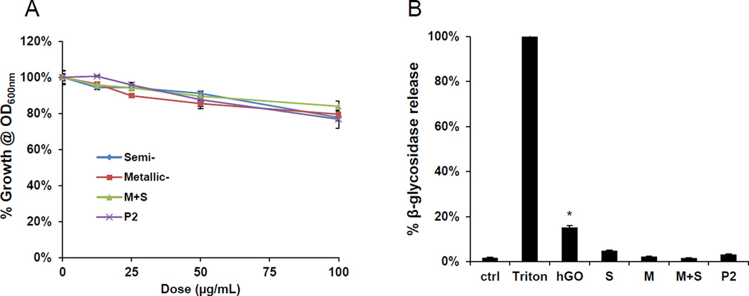
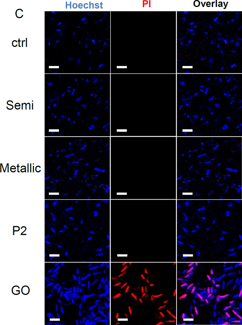
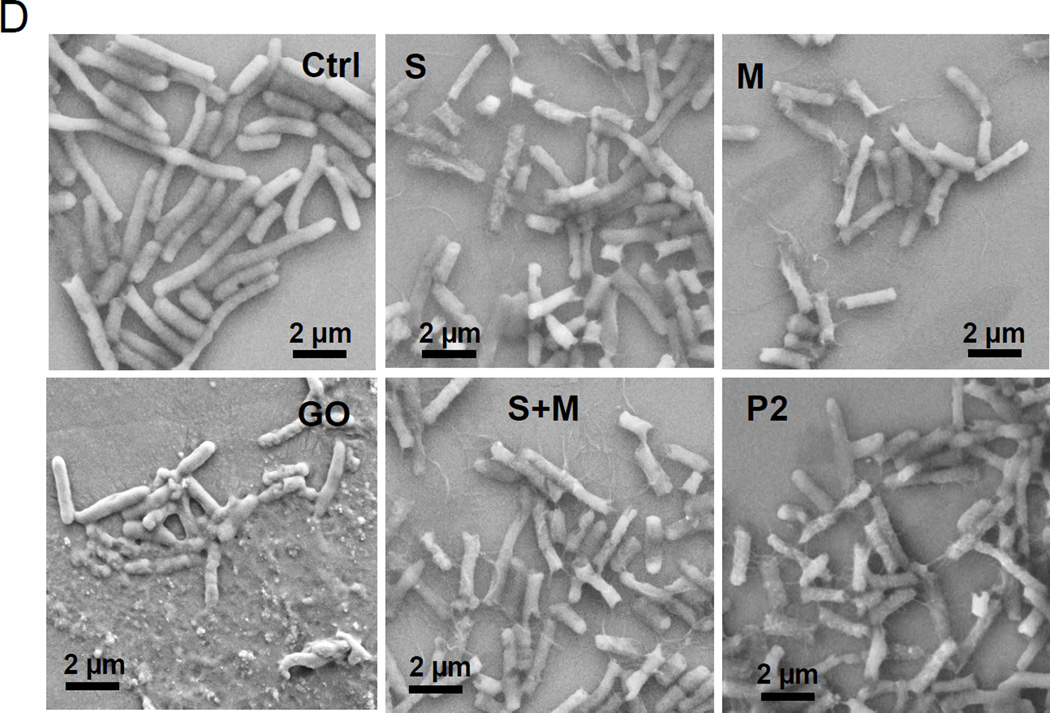
Figure 6. Assessment of the anti-bacterial effects of electronic-sorted SWNCTs in E coli.
(A) Cellular growth was assessed in E coli using a Biotek Synergy plate reader (BioTek, Winooski, VT) at OD600. The cultures were incubated with 12.5, 25, 50 and 100 µg/mL SWNCTs for 24 h. (B) Bacterial cell membrane integrity was evaluated by using a luminescent β-galactosidase substrate to measure the release of the enzyme into the culture supernatant during incubation with 100 µg/mL SWCNTs for 24 h. Triton X-100 membrane lysis was used as a positive control, and for comparative purposes, we also included graphene oxide (GO), which can permeabilize the cell membrane. * p < 0.05 compared to GO treatment. (C) Confocal microscopy to determine bacterial viability during PI staining. Bacteria were seeded into 8-well chamber slides and incubated with 100 µg/mL SWNCTs for 24 h, then the cells were stained with Hoechst 33342 (1 µM) and propidium iodide (5 µM) for 30 min. The cells were washed three times with PBS, and observed under a confocal 1P/FCS inverted microscope. GO was used as a positive control. The scale bars represent 2 µm in length. (D) Scanning electron microscopy (SEM) to determine the morphology change of E. coli. Bacteria grown on a glass substrate were exposed to the SWCNTs at 100 µg/mL for 24 h. After fixation and dehydration, the cells were imaged using a JEOL JSM-67 field emission scanning electron microscope at 10 kV.


