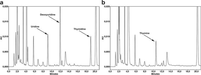
Fig. 2.

Chromatograms of plasma from a MNGIE patient showing thymidine and deoxyuridine peaks. Representative chromatograms obtained from plasma of a MNGIE patient (a), and the same plasma after the selective elimination of TP substrates by treatment with Escherichia coli TP (b). The peaks of dThd and dUrd (5.4 and 10.6 μM in this specific case) virtually disappear in the TP-treated aliquot, whereas the thymine observed in the panel b is the product of dThd phosphorolysis by TP. Uracil, derived from phosphorolyses of dUrd and uridine, is also present, but it elutes very early in the chromatogram and is not resolved well due to coeluting peaks. Note that the ribonucleoside uridine, which is normally found in plasma of healthy controls and MNGIE patients at similar concentrations, is also degraded by E. coli TP.
