Figure 2.
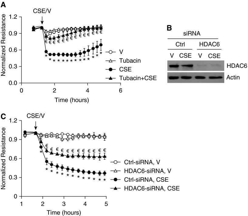
Blocking histone deacetylase (HDAC) 6 blunted CSE-induced EC barrier dysfunction in vitro. (A) Bovine pulmonary artery ECs (PAECs) were preincubated with 100 nM tubacin for 1 hour and then treated with vehicle (V; 15% PBS) or 15% CSE in the absence or presence of 100 nM tubacin for indicated times. Monolayer permeability was assessed by ECIS. (B and C) Human LMVECs were transfected with control siRNA (Ctrl-siRNA) or HDAC6 siRNA (HDAC6-small interfering RNA [siRNA]). At 72 hours after transfection, cells were exposed to vehicle (V; 15% PBS) or 15% CSE for up to 5 hours and monolayer permeability was assessed by ECIS (C). Cells were collected from arrays after ECIS for assessment of HDAC6 protein levels by Western blot (B). Three independent experiments with duplicated ECIS wells for each condition each time were conducted. (B) Representative immunoblots. For A and C, the data are presented as the mean ± SE of the normalized electrical resistance at the indicated time points relative to their initial resistance. ANOVA and Tukey-Kramer post hoc test was used to determine statistically significant difference across means among groups. *P < 0.05 versus V (A) or (Ctrl-siRNA + V) (C); €P < 0.05 versus CSE (A) or (Ctrl-siRNA + CSE) (C). Arrows indicate the time for addition of treatments.

