Figure 2.
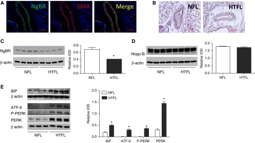
The localization of Nogo-B (NgB) receptor (NgBR) in pulmonary arteries. (A) NgBR localizes to the smooth muscle cell (PASMC) layer of pulmonary arteries in lung section, as indicated by colocalization of NgBR (green) with smooth muscle cell α-actin (red) in pulmonary arteries. (B) NgBR staining intensity is decreased in HTFL pulmonary arteries compared with NFL pulmonary arteries. (C) NgBR protein levels are decreased in HTFL PASMCs compared with NFL twin (n = 4). (D) No difference is observed in the protein levels of ligand NgB between NFL and HTFL PASMCs (n = 4). (E) Markers for endoplasmic reticulum (ER) stress (binding Ig protein [BiP] n = 4; activating transcription factor [ATF]-6, P-protein kinase RNA-like ER kinase [PERK], n = 3) are all increased in HTFL PASMCs. IOD, integrated optic density; SMA, smooth muscle actin. *P < 0.05 compared with NFL. Data are shown as mean (±SE). Scale bars for A: 100 μm and scale bars for B: 200 μm.

