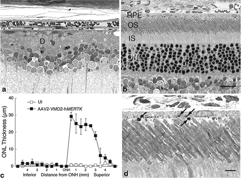
Fig. 65.1.
Structural analysis of RCS rats injected subretinally into one eye with AAV2-VMD2-hMERTK compared with uninjected (UI) contralateral eyes of the same rats, a, b Light micrographs of 1-µm plastic sections of the posterior retina of the UI eye (a), where most photoreceptor nuclei in the ONL have degenerated and disappeared, and an outer segment debris (d) zone is present. The retina of the opposite eye from the eye injected with vector is shown (b), which is comparable in appearance to that of normal rat retinas. c Retinal spidergram showing the ONL thickness along the vertical meridian of UI and vector-injected eyes (each data point is the mean ± SD from 2 rats). d Higher magnification of a vector-injected eye showing phagosomes (arrows) at the apical surface and intracellularly in the RPE. IS inner segments. Scale bars: b = 20 µm; d = 5 µm

