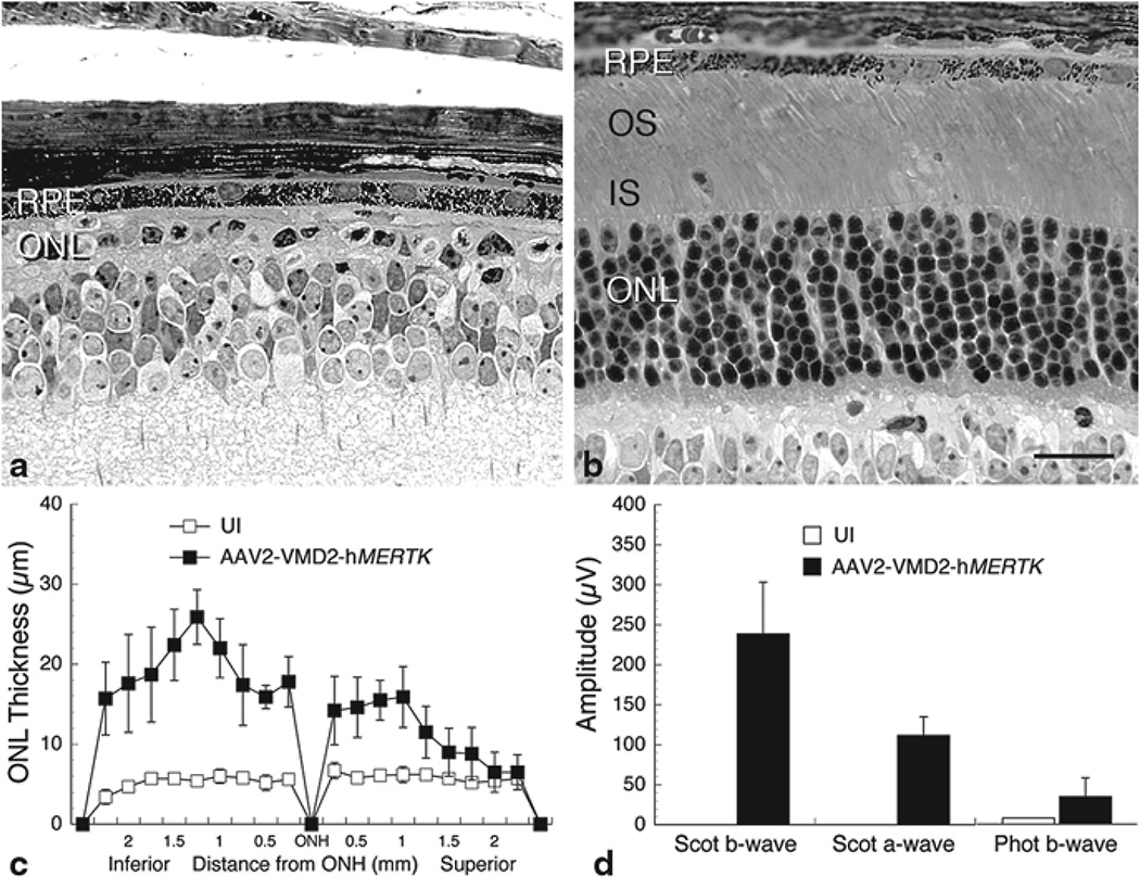
Fig. 65.2.
Structural and functional analysis of Mertk knockout mice injected subretinally into one eye with AAV2-VMD2-hMERTK (b) compared with uninjected (UI) contralateral eyes of the same mice (a). Labeling as described in Fig. 65.1 and in the text. c Retinal spidergram showing the ONL thickness along the vertical meridian of UI and vector-injected eyes (each data point is the mean±SD from 5 mice). d Electroretinographic response amplitudes from the same mice as in c. Scale bar=20 µm

