
Figure 2. Cohesin localization in cohesin mutants and RNAi knockdowns.
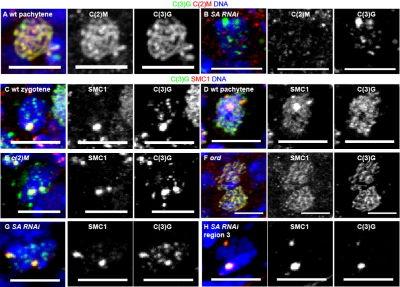
C(2)M (red) localization in (A) wild-type and (B) SA RNAi oocytes with C(3)G in green and DNA in blue. C-H) SMC1 (red) localization and SC assembly with C(3)G (green). The regions of brightest C(3)G and SMC1 are the centromeres (Figure 1) [8, 21, 24]. Zygotene (C) in wild-type is observed in some germaria in the most anterior (earliest) region 2a oocytes. Most of the germarium, region 2a, 2b and 3, contains pachytene (A,D) oocytes. The c(2)M (E) and ord (F) mutant oocytes and SA RNAi (G) oocytes are from the region 2a–2b where pachytene is expected in wild-type. The SA RNAi oocyte in H is from region 3. In all images, the scale bar = 5 μm. See also Figure S2.