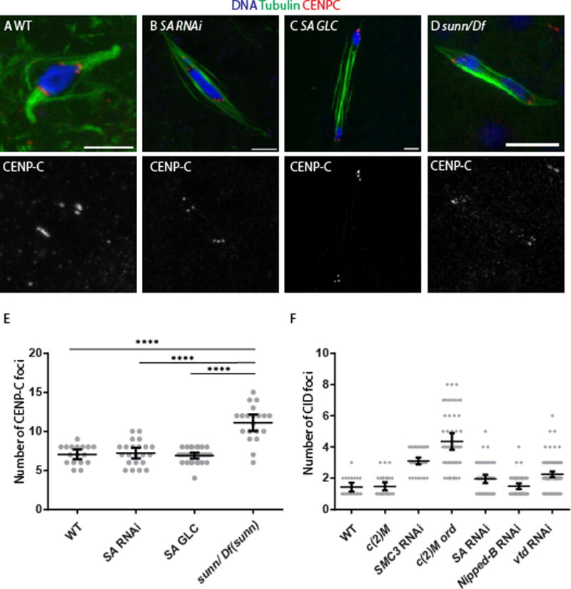
Figure 4. Oocytes lacking SA maintain sister-centromere cohesion.

Mature Stage 14 oocytes, which is where metaphase I occurs, from (A) wild type, (B) SA RNAi, (C) SA germ line clone and (D) a sunn mutant are shown with tubulin in green and CENP-C in red to label the centromeres and the DNA in blue. In some cases, precocious anaphase was observed, which is when the karyosome has separated into two groups of chromosomes that appear to be moving towards the poles. Precocious anaphase was elevated in SA RNAi (60%, n=33), SA germ line clones (15%, n=27) and the sunn mutant (33%, n=30). This phenotype can be caused by a failure in arm cohesion or reduced crossing [29, 47], and because all these mutants or RNAi are expected to reduce crossing over, this phenotype is not necessarily indicative of a cohesion defect. The scale bars are 5 μm. (E) A dot plot showing the number of CENP-C foci in each stage 14 oocyte. Wild-type, and the two SA genotypes are significantly different than sunn, as shown by a Mann-Whitney test. The horizontal and vertical lines show the mean and 95% confidence limits. The number of CENP-C foci expected for wild-type is eight, although less could be observed due to overlap of signals. (F) A dot plot showing the number of CID foci in each pachytene oocyte. The SMC3 RNAi and the c(2)M ord double mutant are significantly different than all the other genotypes (p<0.001) by a Mann-Whitney test. The horizontal and vertical lines show the mean and 95% confidence limits.
