
Fig. 9.
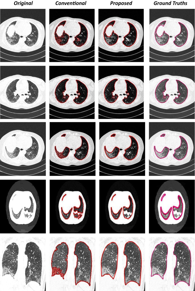


Examples of automated lung region detection results in an axial (1, 2), sagittal (3, 4), and coronal (5) view of the modified convexity algorithm and mathematical morphology technique segmentation for the 12 scans in which an error was detected after the conventional lung segmentation method. The first column shows the original slice, the second column shows the conventional lung segmentation (red contour), the third column shows the modified convexity algorithm and mathematical morphology techniques method (red contour), and the last column shows the result of the manual segmentation (pink contour)