Figure 3.
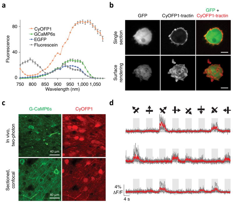
Simultaneous dual-emission two-photon imaging of CyOFP1 with GFP-based reporters. (a) Two- photon excitation spectra of CyOFP1, GCaMP6s, EGFP, and fluorescein (as reference standard). Intensity is presented as thousands of counts per s per μM protein at 1 mW excitation. Error bars are standard deviation. n = 3 for CyOFP1, 4 for GCaMP6s, 2 for EGFP, and 4 for fluorescein. (b) Single optical section (upper row) and surface rendering (lower row) of tractin-CyOFP1 and cytosolic EGFP in MV3 melanoma cells acquired by two-photon Bessel-beam light-sheet microscopy. CyOFP1 is localized to the cortex of the cell and small membrane protrusions. (c) Fluorescence images of CyOFP1 and GCaMP6s in layer-2/3 pyramidal neurons in mouse brain V1 cortex in a single optical section acquired by two-photon excitation at 940 nm. (d) GCaMP6s responses of three mouse neurons co- expressing CyOFP1 in response to drifting gratings. Single sweeps (grey) and averages of 5 sweeps (red) are overlaid. Directions of grating motion (8 directions) are shown above traces (arrows).

