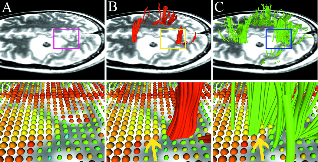
Fig. 1.
Results from case 1 with left frontal metastatic sarcoma. T2-weighted images demonstrate a lesion with significant peritumoral edema and a region-of interest (ROI) is placed on the boundary of the lesion (A) to specify the zoomed-in region (bottom row). Using single-tensor streamline tractography (red fibers), there is no fiber tract that runs through the edematous area (B). Two-tensor unscented Kalman filter (UKF) tractography (green) shows tracing of corticospinal tract (CST) portions that run through the edematous area (C). 3D ellipsoid glyphs show the diffusion tensor model (DTI) in the ROI (D), where isotropic diffusion is shown by red and orange glyph colors. Single-tensor streamline tractography traced fiber tracts only where the tensor ellipsoids are apparently anisotropic (E). In contrast, two-tensor UKF tractography could track in regions with isotropic diffusion (F, arrow).

