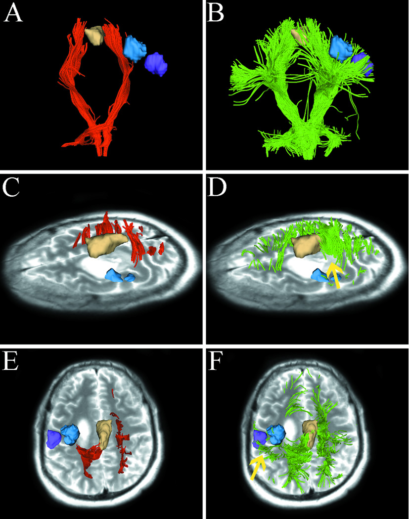
Fig. 3.
Images from case 2 with a left frontal glioblastoma. Images show single-tensor tractography in red, two-tensor UKF tractography in green, and fMRI motor activations of foot (tan), hand (blue), and lip (purple). (A) and (B) show the comparison of tracking results between single-tensor streamline and two-tensor UKF tractography. (C) shows the medial projections of CST which appear to be disrupted because single-tensor streamline tractography underestimates the tracts passing through edema. However, two-tensor UKF tractography delineated some fiber tracts running through the edematous area (D, arrow). The superior views of the results from two different methods are illustrated in (E) and (F), showing that two-tensor UKF tractography better tracked the lateral projections of CST that projected to hand- and lip-related functional cortex (arrow).

