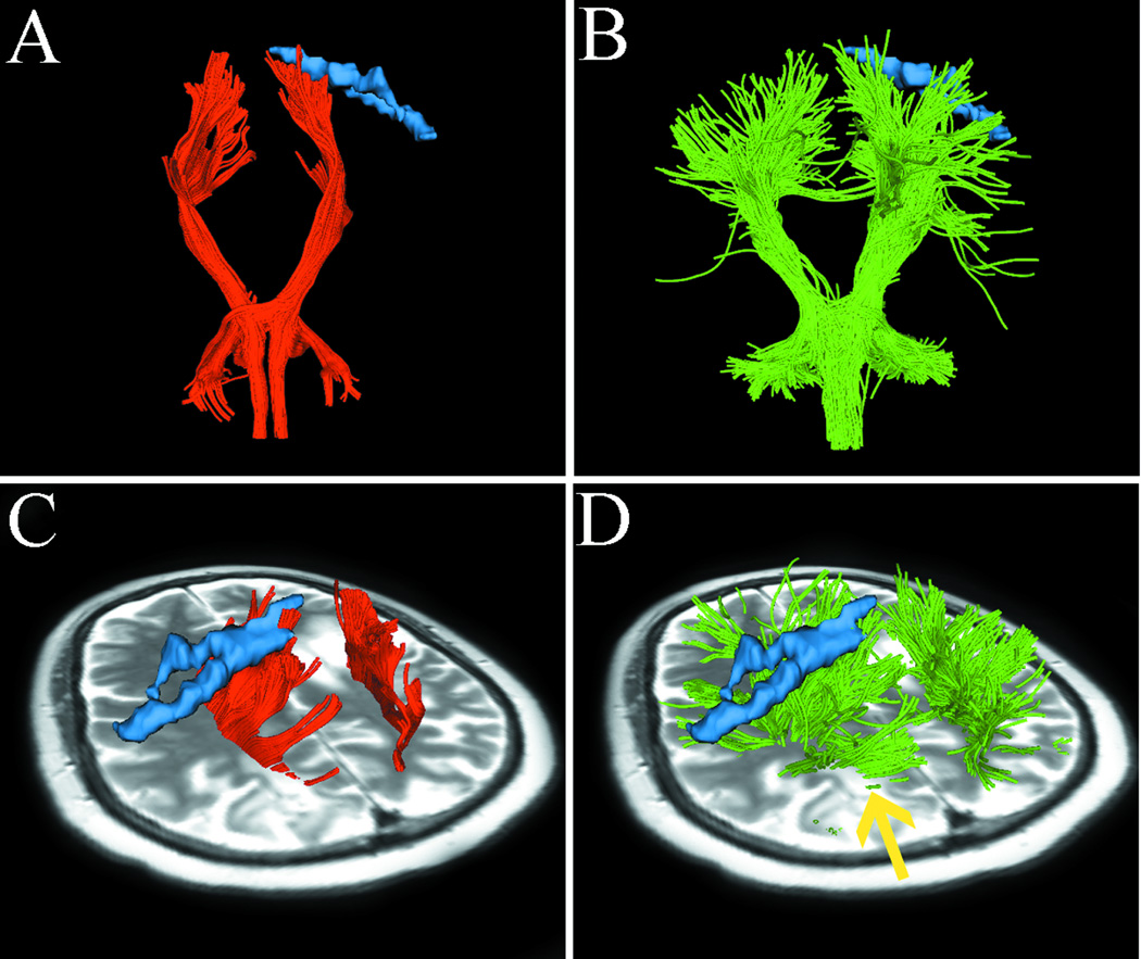
Fig. 5.
Images from case 5 with metastatic breast cancer. Images show single-tensor tractography in red, two-tensor UKF tractography in green, and fMRI motor activations of hand (blue). The lesion of interest for surgical planning and peritumoral edema are indicated by the arrow (D). (A) and (B) show that single-tensor streamline tractography underestimated the tracking of the lateral projections of CST (red), whereas two-tensor UKF tractography traced fanning projections of CST (green). Using the single-tensor streamline algorithm, we found that few fiber tracts linked to hand-related functional cortex and the medial projection appeared to be obscured by the peritumoral edema (C). In contrast, the two-tensor UKF tractography apparently showed the ability to track through regions of edema and the lateral projections of CST which were linked to the hand-related fMRI activation (D, arrow).

