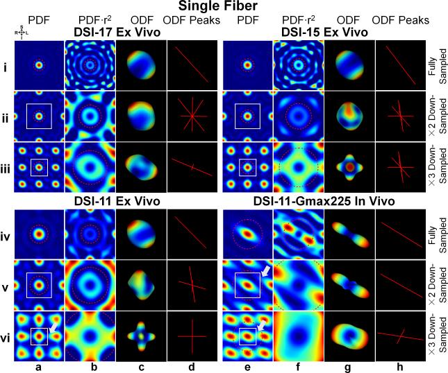
Fig. 7.
The PDF, weighted PDF, ODF and ODF peak for the fully sampled (rows i and iv) and down-sampled (rows ii and v by a factor of 2, rows iii and vi by a factor of 3) q-space signal from a voxel in the corpus callosum of the three ex vivo (DSI-11, DSI-15 and DSI-17) and one in vivo (DSI-11-Gmax225) datasets. Coronal cross-sections through the center of the 3D spin-displacement PDFs are shown in columns a and e. The spatial extent over which the PDF was plotted is kept constant across different down-sampling schemes. The portion of these same PDFs in the field-of-view (FOV, represented by white boxes) is weighted by the square of the displacement distance and shown in columns b and f. All ODFs were reconstructed using unfiltered q-space signal and integrating the PDF to either the estimated mean displacement distance (MDD, red dashed circles) or the new FOV after down-sampling if it is smaller than MDD (i.e. row vi, column b; rows v and vi, column f). The ODFs that correspond to the PDFs in columns b and f are shown in columns c and g. White arrows indicate the regions of aliasing artifacts in the PDF. The color map represents low values (blue) to high values (red) in both the PDF and ODF.

