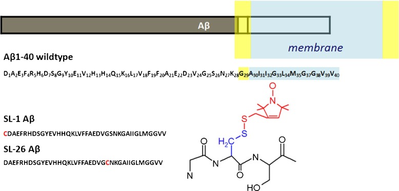
Fig. 1.
Overview of A β1-40 sequence and constructs. Top: Schematic of A β1–40 relative to membrane location of the amyloid precursor protein (APP) [3, 5–9], membrane dimensions not to scale. Light blue: hydrophobic part of the membrane, yellow: lipid headgroup region. Middle: A β1–40 sequence, bottom left: constructs used. Red: cysteine used to link MTSL spin label. Bottom right: MTSL-linked to a schematic protein backbone

