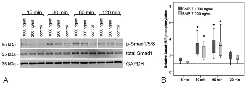
Figure 3. Phosporylation of Smad1/5/8 after BMP-7 stimulation.
(A) Exemplary western blots show increased Smad 1/5/8 phosphorylation after 30 and 60 min of stimulation with BMP-7, but no increase in total Smad1 levels compared to unstimulated controls. GAPDH serves as reference protein. Imaging was conducted using the Odyssey imager and LiCor Odyssey software. (B) Quantification of relative phosphorylation normalised to total Smad1 and GAPDH was significantly increased at 30 and 60 min (n = 6). Stars mark significant differences to the untreated control, p = 0.002.

