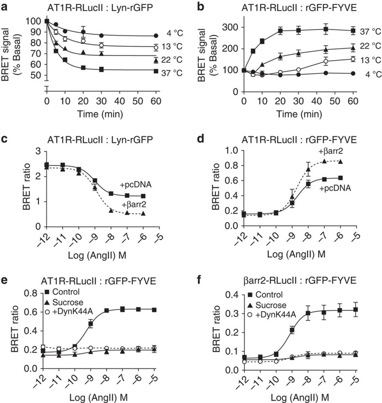
Figure 3. AngII-mediated AT1R and β-arrestin2 trafficking assessed by EbBRET.
(a,b) HEK293 cells were transfected with AT1R-RlucII along with either Lyn-rGFP (a) or rGFP-FYVE (b). Cells were incubated at 4 °C, 13 °C, 22 °C (RT) or 37 °C for the indicated times in the presence of AngII (100 nM) before BRET measurements. The BRET signals are expressed as a percentage of BRET ratios observed in the control (without AngII treatment). Data shown represent the means±s.e.m. of three independent experiments. (c,d) HEK293 cells were transfected with AT1R-RLucII/Lyn-rGFP (c) or AT1R-RlucII/rGFP-FYVE (d) along with either pcDNA or β-arrestin2. Cells were treated with the indicated concentrations of AngII for 30 min (c) or 40 min (d) before BRET measurements. Raw BRET ratios are the means±s.e.m. from at least three independent experiments. (e,f) HEK293 cells were transfected with AT1R-RlucII/rGFP-FYVE (e) or AT1R/βarr2-RlucII/rGFP-FYVE (f), along with either pcDNA or dynamin K44A (DynK44A, O). Cells were incubated in the absence (control, ▪) or in presence of 0.45 M sucrose (▴) for 20 min, and stimulated with the indicated concentrations of AngII for 40 min before BRET measurement. Raw BRET values are the means±s.e.m. of three independent experiments.

