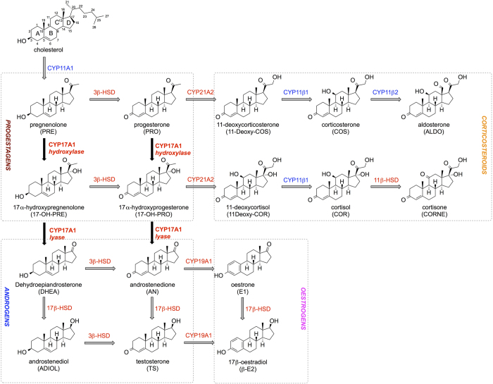
Figure 1. Summary of the steroidogenesis process.
Enzymes coloured in blue are located in the mitochondrial membrane, while the red ones are present in the smooth endoplasmic reticulum. Reactions catalysed by CYP17A1 are reported in bold and black arrows. Abbreviations for each steroid are reported in brackets. Other abbreviations: HSD (hydroxysteroid dehydrogenase).

