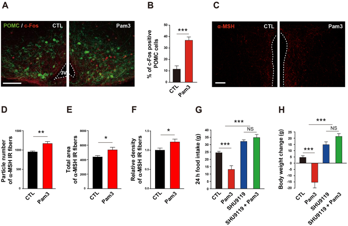
Figure 6. Involvement of melanocortin pathway in the TLR2-induced anorexia and body weight loss.
(A,B) POMC neuronal activity was determined by the change in c-Fos immunoreactivity in the arcuate nucleus of mice that were icv treated with Pam3CSK4 (Pam3) 1 h before sacrifice. Representative photographs (A) and calculated graphs (B) reveal an increase in the number of c-Fos positive POMC cells induced by Pam3 (CTL, n = 4 section/2 mice; Pam3, n = 6 section/3 mice; ***P < 0.0001 by unpaired two-tailed Student’s t-tests). Scale bar = 100 μm. (C–F) Effect of Pam3 on POMC neuronal terminals of the hypothalamic paraventricular nucleus (PVN) was determined by α-melanocyte stimulating hormone (α-MSH) immunoreactivity. Representative images (C) show an increase in α-MSH immunosignals in the PVN after Pam3 injection. Scale bar = 100 μm. Icv administration of Pam3 led an increase in particle number (D), total area (E), and relative density (F) of α-MSH fiber signals (CTL, n = 12 sections/6 mice; Pam3, n = 16 sections/8 mice; *P < 0.05, **P < 0.01 by unpaired two-tailed Student’s t-tests). (G,H) Effect of antagonizing melanocortin 3 and 4 (MC3/4) receptors on Pam3-induced sickness behaviors. Changes in food intake and body weight were monitored in rats after icv treatment with SHU9119, an MC3/4 receptor antagonist. The antagonist completely abolished the inhibitory effect of Pam3 on food intake (G) and body weight (H) (n = 4–5 rats/group; ***P < 0.001 by two-way ANOVA; NS, not significant). (G,H) Data from rat model. All data are presented as mean ± s.e.m.

