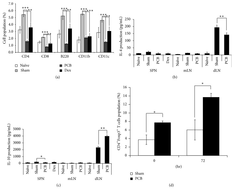
Figure 3.
PCB extract suppressed infiltrated immune cell and Th2-related cytokine via Foxp3 regulation in a mouse model of AD. (a) For analyzed immune cells in ear, ear tissues were isolated single cells. The cells were stained with with anti-CD4 (FITC), anti-CD8 (APC/Cy7), anti-CD11c (PE), anti-CD11b (APC), and anti CD45R (PerCP/Cy5.5). These surface molecules of immune cells were measured by flow cytometry. To measure cytokines, the cells isolated from dLNs were restimulated with 100 μg/mL HDM extract and cultured for 72 h. IL-4 (b) and IL-10 (c) were detected by ELISA. (d) To evaluate population of CD4+ Foxp3+ T cells, the cells isolated from dLNs were cultured with PMA (50 ng/mL) and Ionomycin (250 ng/mL). After staining with CD4 and Foxp3, the population of CD4+ Foxp3+ T cells was analyzed by flow cytometry at 0 h and 72 h. Each value is shown as mean ± SD (n = 3). ∗ P < 0.05, ∗∗ P < 0.01, and ∗∗∗ P < 0.001 compared with sham group. Data were analyzed using ANOVA followed by F-protected Fisher's least significant difference test.

