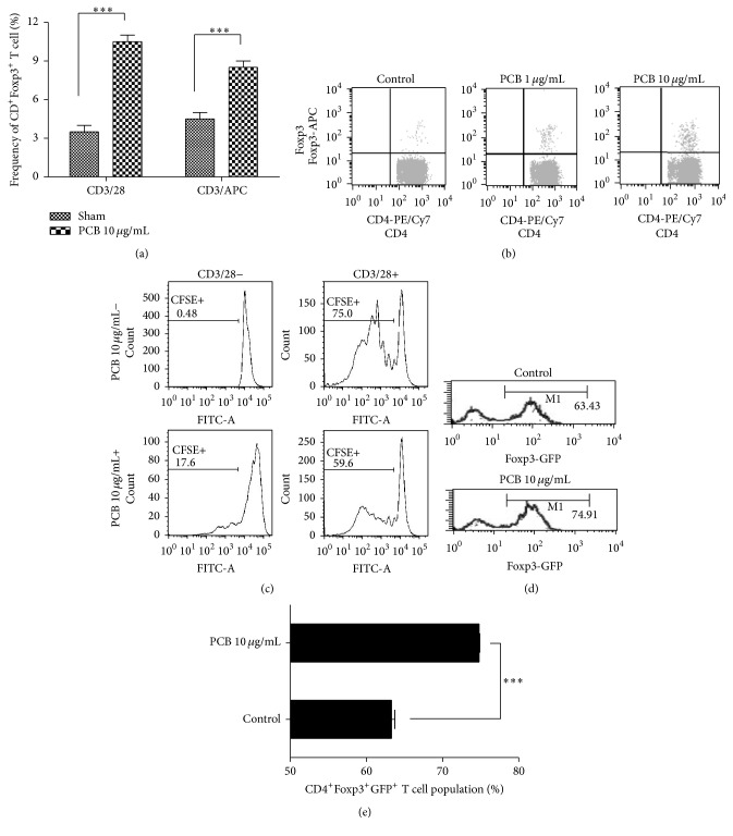
Figure 6.
PCB extract affects the generation and functional Foxp3+ T regulatory cells and stabilizes the Foxp3 expression of regulatory T cells. (a) and (b) CD4+CD62L+ naïve T cell from splenocytes and mLN cells from BALB/c mice were cultured with 10 μg/mL plate-bound anti-CD3 monoclonal antibody and 2 μg/mL soluble anti-CD28 mAb, or with 10 μg/mL plate-bound anti-CD3 mAb and APCs, in the presence of 0–10 μg/mL PCB. After three days, the expression of Foxp3 in gated CD4+ cells was analyzed using flow cytometry. A plot from one representative experiment shows the frequency of Foxp3+CD4+T cells. (c) CD4+ T cells from each group were cocultured with CD4+CD62L+ naïve T cells labeled with CFSE (5 μM), 10 μg/mL plate-bound anti-CD3 mAb, and 2 μg/mL soluble anti-CD28 mAb. The CFSE+ population was then analyzed using FACS. (d) Ag-specific GFP+ nTregs were sorted from the OT-II transgenic Foxp3-GFP knock-in mice and transferred to normal recipients. Five days after immunization, the transferred CD45.2+ cells were analyzed for Foxp3 expression using flow cytometry. (e) The numbers in M1 represent the percentage of Foxp3+ cells. The plot presents the mean percentage of CD45.2+Foxp3+ T cells of three independent experiments. Data are presented as the mean ± SD of triplicate determinations and were analyzed using unpaired t-test. ∗∗∗ P < 0.001 versus control.

