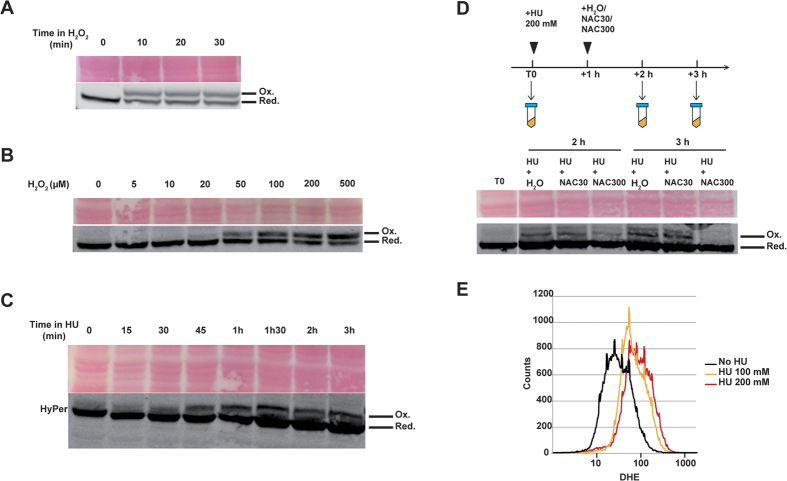
Figure 4. Hydrogen peroxide is detected in yeast cells after hydroxyurea exposure.
(A–D) HyPer detection using redox western blotting. (A) Exponentially growing yeast cells expressing HyPer cultured in YPD were exposed to H2O2 (1 mM) for increasing times as indicated. Oxidized (Ox.) and reduced (Red.) HyPer bands are indicated, and Ponceau red staining is shown as loading control. (B) Yeast cells were treated for 20 min with increasing H2O2 concentrations as indicated. (C) Yeast cells were treated with HU (200 mM) for increasing times as indicated. (D) Yeast cells were treated with HU (200 mM) for 1 hour. NAC (30 mM or 300 mM) was then added to cultures for 1 hour and 2 hours. H2O was also used as a control. Samples were processed at various times as indicated. (E) ROS detection using DHE. Exponentially growing yeast cells cultured in YPD were exposed to HU (100 and 200 mM) for 1 hour, before staining with DHE to evidence ROS production.

