
Figure 8. Identification of a Treg-stimulating ppins76–90 peptide.
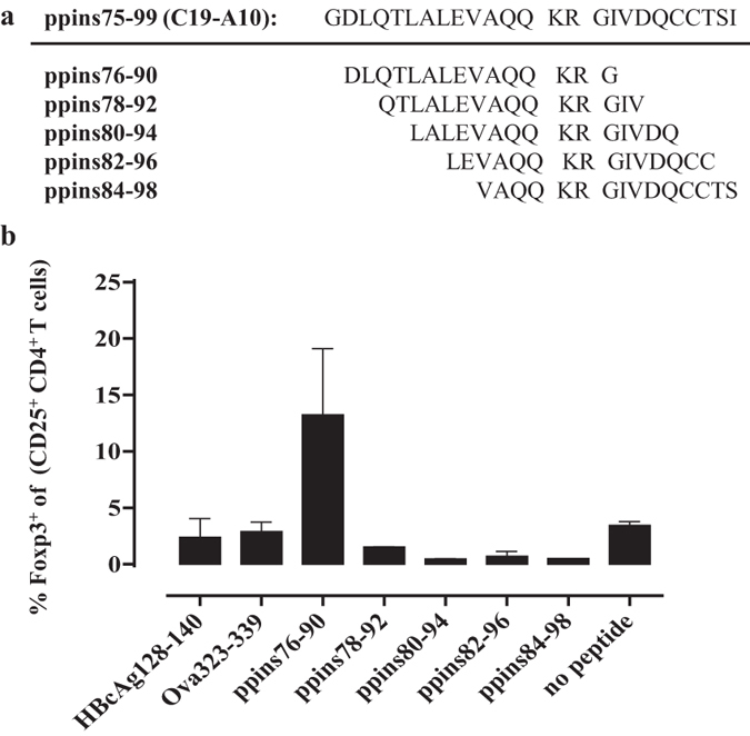
(a) Aminoacid sequences of the ppins75–99 (C19-A1) fragment and overlapping 15-residue peptides ppins76–90, ppins78–92, ppins80–94, ppins82–96 and ppins84–98. (b) Conventional Foxp3negative/eGFPnegative CD4+ T cells were isolated from spleens of B6-Foxp3eGFP mice using magnetic assisted (MACS) and fluorescence assisted (FACS) cell sorting and stimulated for three days with CD3-depleted autologous splenocytes pulsed with the respective ppins-derived peptides as well as two I-Ab-binding control peptides (HBcAg128-140 and Ova323-339). In vitro conversion of Foxp3negative/eGFPnegative CD4+ T cells into Foxp3+/eGFP+ CD25+ CD4+ T cells was determined by FCM (see Supplementary Protocols). The actual percentages of newly arising Foxp3 expressing CD25+ CD4+ Treg cells ± SD of a representative experiment (out of two experiments performed) are shown.