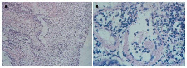
Figure 1.

Chronic radiation proctitis. A: Chronic radiation proctitis (low power view). This picture shows the mucosa with severe oedema, non-specific inflammation, lymphocytosis, hyalinization in the stroma and fibrin thrombi in the postcapillary venules (Hematoxylin and Eosin stain, 10 ×); B: Chronic radiation proctitis (High power view). This picture shows two veins in the lamina propria, one with patchy occlusive fibrin thrombus. The wall shows thickening and hyalinization. A dense non-specific inflammation including few eosinophils also seen (Hematoxylin and Eosin stain, 40 ×).
