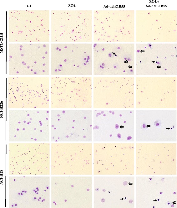
Fig. 4.

Giemsa staining after Ad-delE1B55 infection. Cells were treated with ZOL (MSTO-211H: 10 μM, NCI-H226: 60 μM, NCI-H28: 80 μM) and/or Ad-delE1B55 (MSTO-211H: 2 × 10³ vp/cell, NCI-H226: 1 × 10³ vp/cell, NCI-H28: 2 × 10³ vp/cell) for 72 h. Nuclear morphological changes were examined after the Giemsa staining. In Ad-delE1B55-treated cells, small and highly condensed nucleus (solid arrow) and large and uncondensed nucleus (open arrow) were detected. An upper panel and lower panel are x4 and x10 in magnifications, respectively
