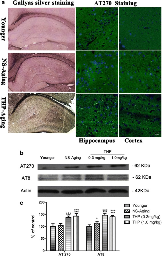
Fig. 6.

Qualitative and quantitative profiles of neurofibrillary pathology in rats after long-term THP treatment. a Neurofibrillary tangles stained by Gallyas silver staining in the hippocampus and immunolabeling with antibody AT270 recognizing tau phosphorylated at Thr181. Gallyas silver staining revealed more silver-positive staining with a enhanced expression profile of AT270-positive neurofibrillary tangles in the brain of rats with long-term THP exposure. b Neurofibrillary pathology was quantified by western blot analysis in the prefrontal cortex as a brain region of interest. c Qualitative analysis revealed significantly upregulated AT8 and AT270 expression in THP-treated aging rats compared with the NS-aging group. *P < 0.05, ***P < 0.0001 compared with the younger group (3 months old); &P < 0.05, &&&P < 0.0001 compared with the NS-treated group (Dunnett’s test). Scale bars = 20 μm
