Fig. 7.

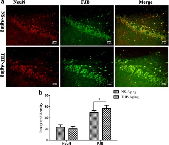
Confocal microscopy revealed hippocampal neurodegeneration in rats after long-term THP treatment. Hippocampal neuronal loss and neurodegeneration were detected using NeuN and FJB, fluorescent markers for neurons and neurodegeneration, respectively. a Confocal microscopy shows colocalization of some neurons with neurodegeneration. Scale bar = 50 μm. b Integrated immunofluorescence density analysis was performed using ImageJ. THP-treated rats show slightly increased FJB-labeled cells in selected hippocampal areas. * P < 0.05 compared with the NS-treated aging controls (Student’s t test)
