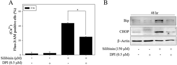
Fig. 5.

Mitochondrial ROS mediated silibinin-induced ER stress in PC-3 cells. a PC-3 cells were treated with 150 μM silibinin for 24 h with the presence or absence of 0.5 μM DPI. The intracellular Ca2+ concentration was determined by the fluorescence of fluo-3/AM. b The effects of DPI on silibinin-induced ER stress were measured by western blotting. β-Actin was used as a loading control. Data are presented as mean ± SD (n = 3 in each group). * p <0.001 vs. the control group.
