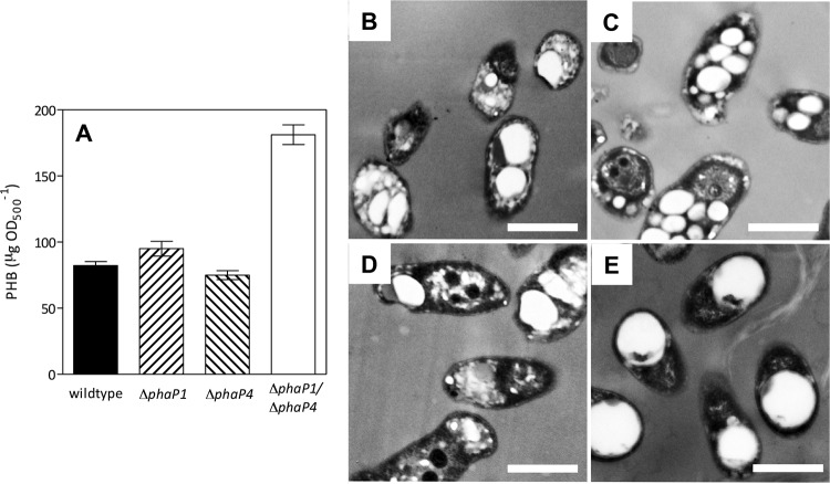
FIG 3.
PHB accumulation in the wild type and three ΔphaP mutant derivatives, evaluated at 17 days under oxic conditions. (A) PHB extracted from cells, expressed as the average from four independent determinations ± SEM. Transmission electron micrographs of the wild-type (B), ΔphaP1 (C), ΔphaP4 (D), and ΔphaP1 ΔphaP4 (E) strains. Note the PHB granules as bright spots inside the cytoplasm. Scale bars, 0.5 μm.

