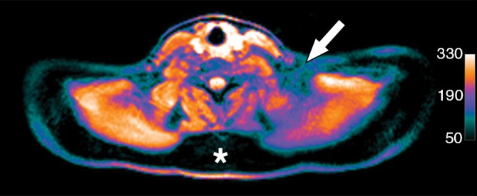
Figure 5e:

Images in healthy 22-year-old female volunteer (body mass index: 28 kg/m2) after cold activation of BAT by using a cooling vest protocol. (a) Coronal Dixon fat-only MR image for anatomic reference shows cervical and supraclavicular fat (arrow). (b) Fused 18F-FDG PET/MR image shows increased FDG uptake in areas corresponding to fat (arrow), consistent with BAT. (c) Axial Dixon fat-only MR image for anatomic reference. (d) Fused 18F-FDG PET/MR image shows increased FDG uptake in supraclavicular (white arrow) and intermuscular (black arrow) fat consistent with BAT. (e) Axial Dixon water-only MR image at same level shows pixel values indicating relatively higher water content in FDG-avid supraclavicular fat (arrow) compared with subcutaneous fat (*).
