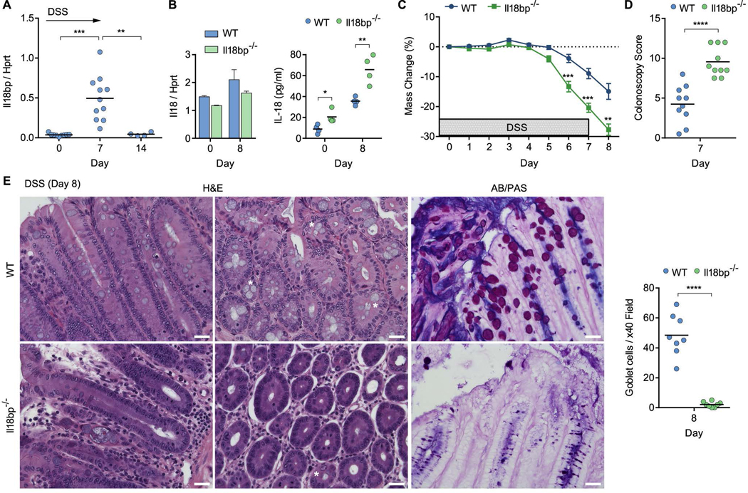
Figure 3. Hyperactive IL-18 signaling drives colitis and goblet cell loss in Il18bp−/− mice.
(A) Wild-type (WT) mice were treated with 2% DSS in drinking water and Il18bp mRNA expression in the distal colon was measured over 14 days. (B) IL-18 mRNA expression in distal colon (left) and protein secretion in colonic explants (right) of il18bp−/− and WT littermates. (C) Weight loss following DSS treatment in cohoused WT and Il18bp−/− littermates (n=10). (D) Colonoscopy severity score of WT and Il18bp−/− mice. (E) Representative H&E and AB/PAS staining of distal colon sections obtained from cohoused WT (top) and Il18bp−/− (bottom) littermates on day 8 post DSS treatment. Asterisks mark example goblet cells. Right, enumeration of goblet cells in histological sections from cohoused WT and Il18bp−/− littermates. Scale bar = 25 μm. Data are represented as mean ± SEM. *, p<0.05; **, p<0.01; ***, p<0.001; ****, p<0.0001 by unpaired Student’s t-test. Related to Figure S1–4.

