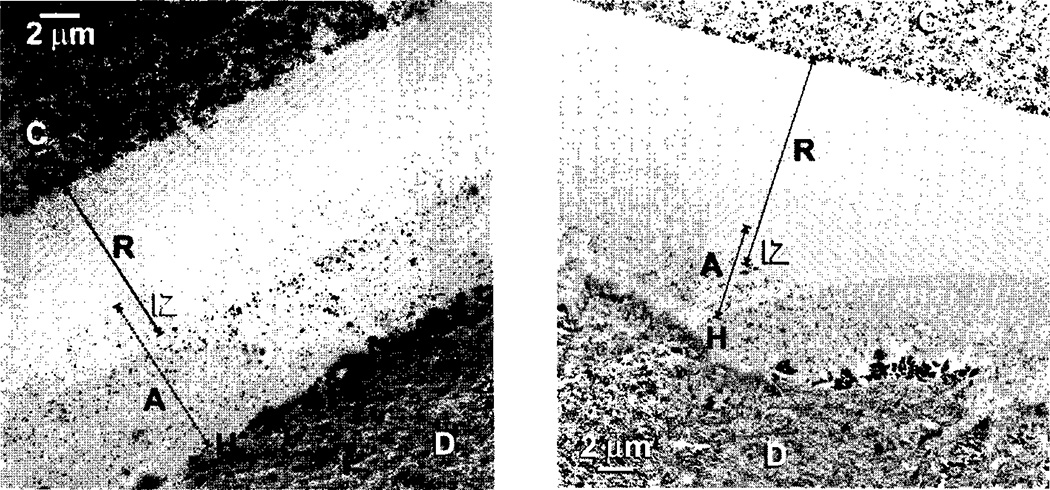
Fig. 10.
TEM micrographs of (A) dentin bonded with a coating of iBondi, followed by solvent evaporation and light-curing. The bonded surface was then coated with Scotchbond Multi-Purposea adhesive that was light-cured. After soaking in 50% ammoniacal silver nitrate overnight, the specimens were processed for transmission electron microscopy (TEM). Note the moderate silver nanoleakage in the bottom layer of iBondi adhesive (A) and the near absence of nanoleakage in the SBMPa adhesive (R). (B) Dentin bonded with one layer of Xeno IIIf, then the solvent was evaporated and the surface light-cured. The bonded surface was covered with one coat of water-free SBMPa adhesive (R). Note there is much less silver nanoleakage in (R) than in (A) (from Ito et al.52, with permission).

