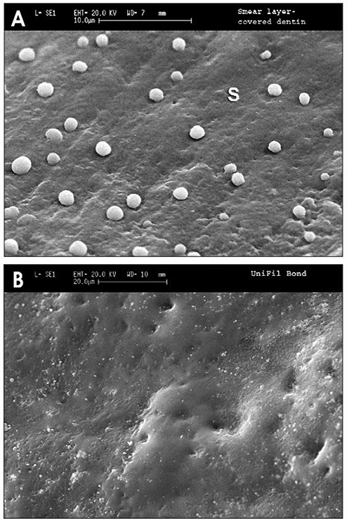
Fig. 3.
SEMs of resin replicas of crown preparations of vital teeth covered with (A) smear layer. Note presence of a few microdroplets of transudated dentinal fluid trapped by the impression material. (B) Unifil Bondd bonded surface after removal of O2-inhibited resin. No fluid droplets were seen when using this two bottle, self-etching primer adhesive (from Chersoni et al.36, with permission).

