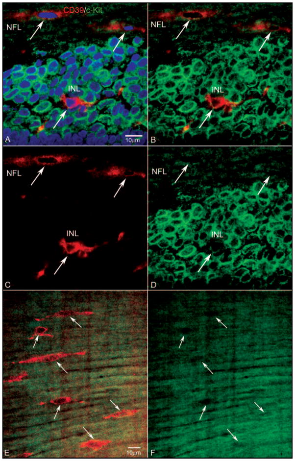
Figure 3.
Expression of CD39 and c-Kit in a retinal cross-section (A–D) and a retinal wholemount (E, F) from a 7-WG embryo. Blue: DAPI-counterstained nuclei; red: CD39; green: c-Kit. (A) Near the nerve head, fusiform cells in the NFL were CD39+/c-Kit−, whereas some cells in the inner neuroblastic layer (INL), were CD39+/c-Kit+ (A–D, arrow in INL). (B) Same section without the DAPI channel, showing that c-Kit+ cells occupied the entire region along with some double-labeled cells. The fusiform cells in the NFL appeared to be c-Kit−. (C) The same section showing only the CD39 channel. The CD39+ cell (arrow) in the INL had processes, suggesting it was migrating. (D) Same section showing only the c-Kit channel. The CD39+ cell in the INL appeared to be less c-Kit+ than did the c-Kit+/CD39− neighboring cells. (E) Retinal wholemount showing CD39+ fusiform cells near the nerve head (arrows) aligned with very linear cells expressing c-Kit. (F) Same region as shown in (E), with only the c-Kit channel showing that the cell expressing CD39 were negative for c-Kit. Scale bars: (A) applies to (B–D); (E) applies to (F).

