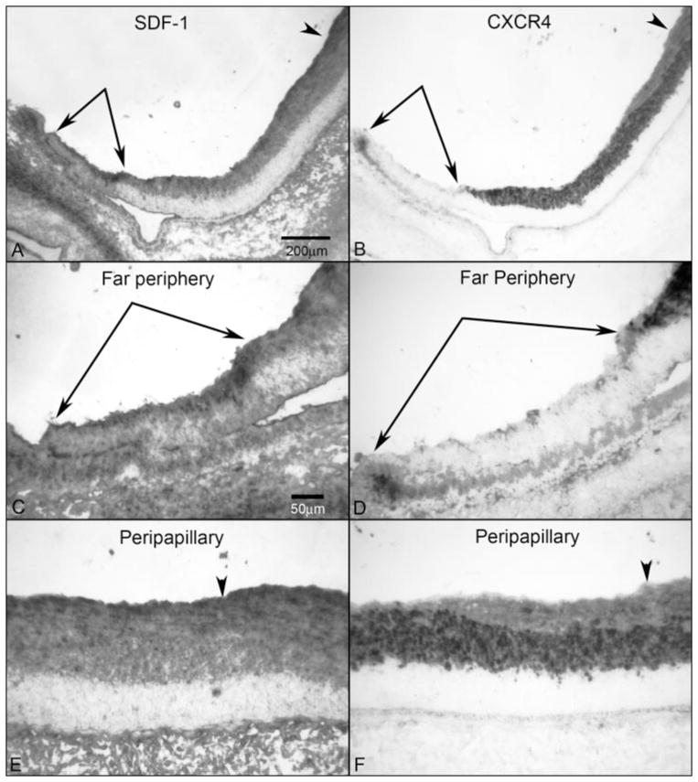
Figure 4.
SDF-1/CXCR4 axis in a 12-WG fetal eye. Low-magnification micrographs showing SDF-1 (A) and CXCR4 (B) from the peripapillary region adjacent to the optic nerve head (arrowhead) to the far periphery (paired arrows). In the far periphery, SDF-1 was more intense in the region where CXCR4+ precursors had not yet migrated (paired arrows). (C, D) Higher magnification of the peripheral retina showing more intense SDF-1 in advance of (paired arrows) CXCR4+ precursors. (E, F) Higher magnification of the peripapillary region showing less intense SDF-1 immunoreactivity than in the far periphery. CXCR4 immunoreactivity was present throughout the NFL at this age but was less intense than in the INL. Arrowhead: ILM. Stain: APase/NBT reaction product after bleaching. Scale bar: (A) applies to (B); (C) applies to (D–F).

