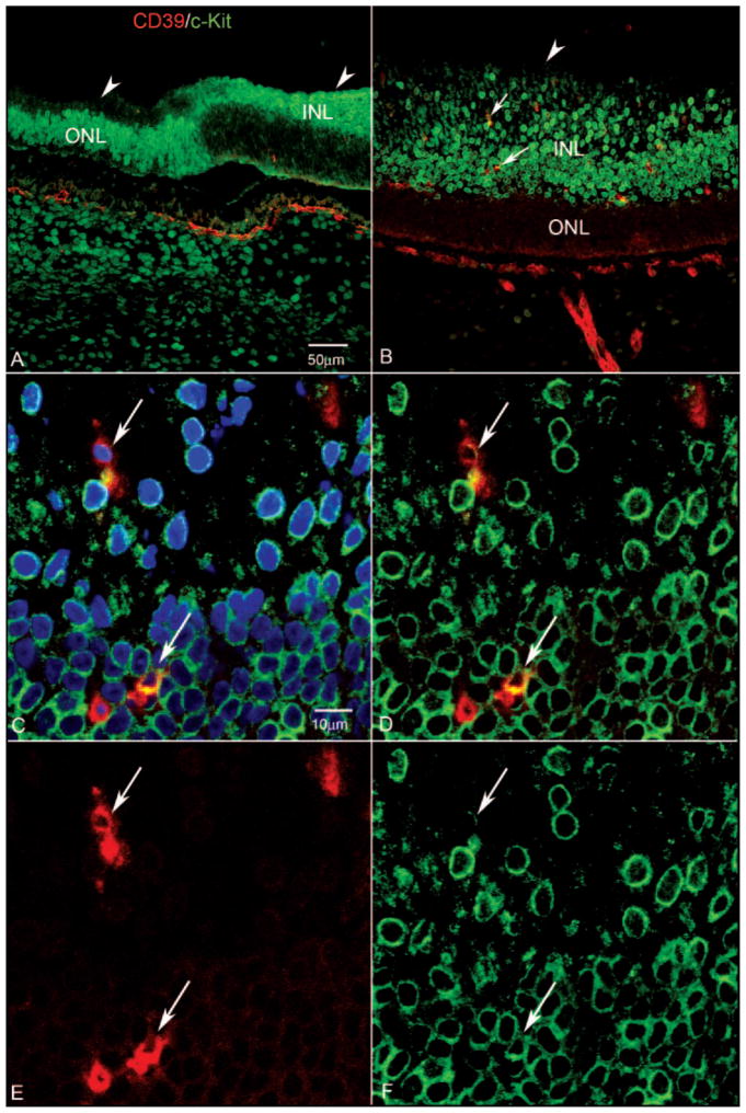
Figure 6.
Expression of CD39 and c-Kit in retinal cross section of a 12-WG fetal eye at low (A, B) and high (C–F) magnifications. Blue: DAPI-counterstained nuclei; red: CD39; green: c-Kit. (A) In the far periphery in this region of retina, c-Kit-expressing cells were present in the inner neuroblastic layer (INL) posteriorly and in the outer neuroblastic layer (ONL) more peripherally. Arrowheads: ILM. Very few CD39+ cells were evident in this region at this age. (B) Near nerve head, c-Kit+ cells occupied the entire extent of the INL. Some of these cells expressed CD39 (arrows). Arrowhead: ILM. (C) Higher magnification of the same cells indicated with the arrows in (B) showing that they were CD39+/c-Kit+ (arrows). (C–F) Arrows: same cells. (D) Same section without the blue DAPI channel, showing c-Kit-immunostained cells occupying the entire INL and some double-labeled cells in this region. (E) Same section showing only the CD39 channel. (F) Same section showing only the c-Kit channel. The CD39+ cell closest to the NFL (top arrow) appeared to have less c-Kit than did the deeper lying CD39+ cell. Scale bars: (A) applies to (B); (C) applies to (D–F).

