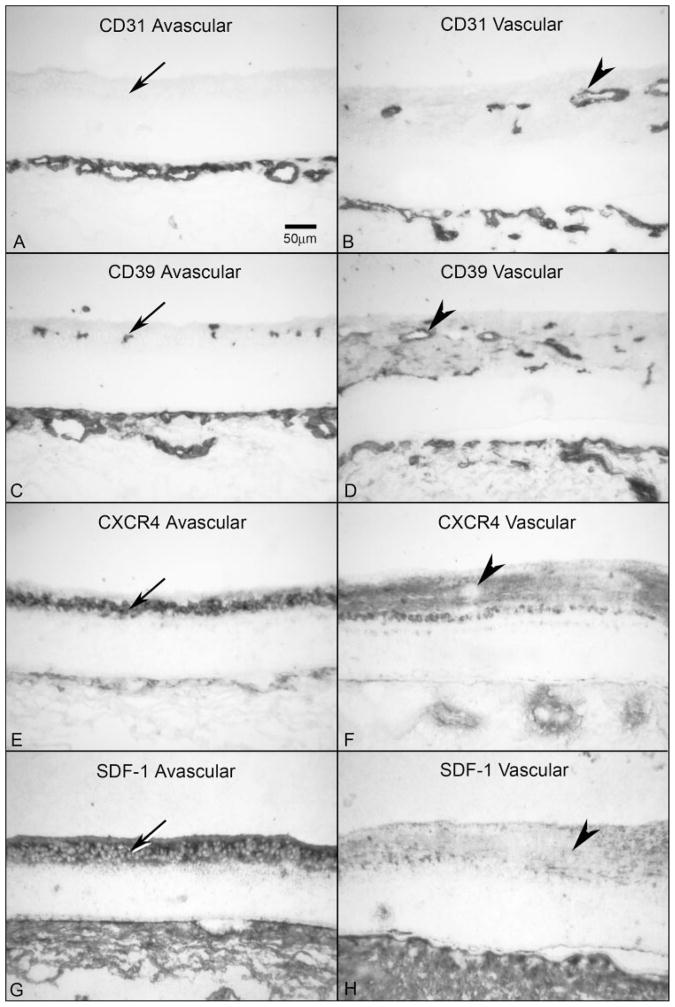
Figure 7.
Comparison of CXCR4 and SDF-1 localization in avascular and vascularized retina from a 16-WG fetal eye. (A, B) CD31 was absent in avascular peripheral retina (A, arrow) but clearly labeled blood vessels in vascularized regions (B, arrowhead). The choriocapillaris is the layer of CD31+ blood vessels at the bottom in both panels. (C, D) CD39 was present on angioblasts in avascular retina (C, arrow) and in blood vessels and angioblasts in vascularized areas (D, arrowhead). (E, F) In avascular retina, CXCR4 labeling was intense in angioblasts within the PGCL (E, arrow), but was significantly reduced in areas that are vascularized and absent in retinal vessels (F, arrowhead). (G, H) SDF-1 immunoreactivity was much more intense in avascular inner retina (G, arrow) than in vascularized retina (H, arrowhead). Stain, APase/NBT blue reaction product. Scale bar: (A) applies to all panels.

