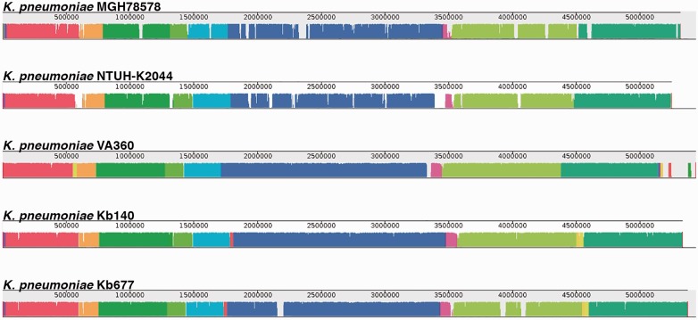
Fig. 4.—
Alignment of K. pneumoniae strains genomes using MAUVE. Each genome’s panel contains a scale showing the sequence coordinates, colored blocks that represent regions of the genome sequence that aligned to part of the other genomes, and a single black horizontal center line where the blocks that lie above are in the forward orientation (in this case there are no blocks in reverse orientation, then the lines look to be at the bottom across the genome).

