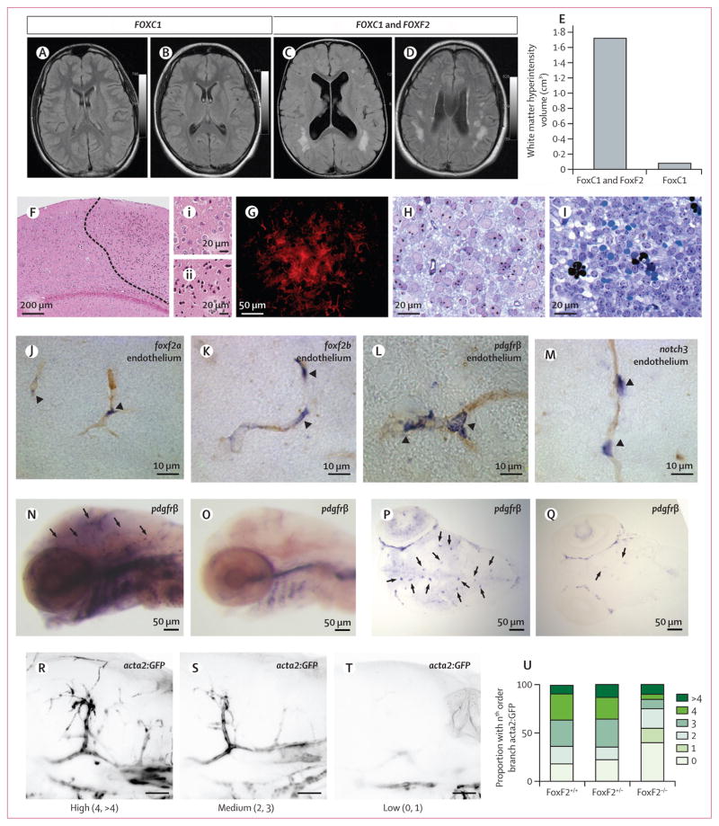
Figure 2. Cerebrovascular phenotype of FOXF2 and FOXC1 deletions in humans and FoxF2 knockout mice, and expression of foxf2a and foxf2b in zebrafish.
(A–D) In two patients with segmental deletion encompassing FOXC1, white matter hyperintensities are noted in periventricular region (A,B) and subcortical regions (B). In two patients with segmental deletion of both FOXC1 and FOXF2 (C,D), mean white matter hyperintensities volume is increased by more than ten times (E), in subcortical and periventricular regions (see appendix p 68 for white matter hyperintensities volumes in each of the four patients). (F–I) Cerebral cortex of conditional Foxf2 knockout mouse showing ischaemic infarction and haemorrhagic tissue. (F) Area with condensed eosinophilic cytoplasm and pyknotic nuclei (to the right dashed line) that indicates recent ischaemic infarction. (Fi) Normal tissue and (Fii) tissue with ischaemic infarction at higher magnification. (G) Glial fibrillary acidic protein immunofluorescence of area with reactive astrogliosis in cerebral cortex of Foxf2 conditional knockout mouse. (H) Cerebral cortex from control mouse showing normal neuronal tissue and intact capillaries. (I) Haemorrhagic area of cerebral cortex from Foxf2 conditional knockout mouse. Extravascular erythrocytes seen both as intact cells (homogeneous greenish blue) and lysed cells (black). (J–M) RNA in situ hybridisation (purple) of larval zebrafish brains shows expression of foxf2a (J) and foxf2b (K) in presumptive pericytes compared with known pericyte markers pdgfrβ (L) and notch3 (M; purple) around capillaries in 1-month old larval zebrafish (brown). (N-Q) foxf2−/− mutants have reduced expression of pericyte marker pdgfrβ in 4-day postfertilisation embryonic cerebral vasculature. (R–U) Loss of foxf2b results in reduction of the smooth-muscle marker acta2:GFP coverage of blood vessels in wild type (n=11), foxf2+/− (n=31), and Foxf2−/− (n=20) embryonic cerebral vasculature. (R–T) Examples of high, medium, and low branch order coverage scored from 0 to the 4th order branch in vessel coverage presented as percentages of total embryo counts (U). Arrow heads in images J–Q refer to positions showing pericytes.

