
Figure 1.
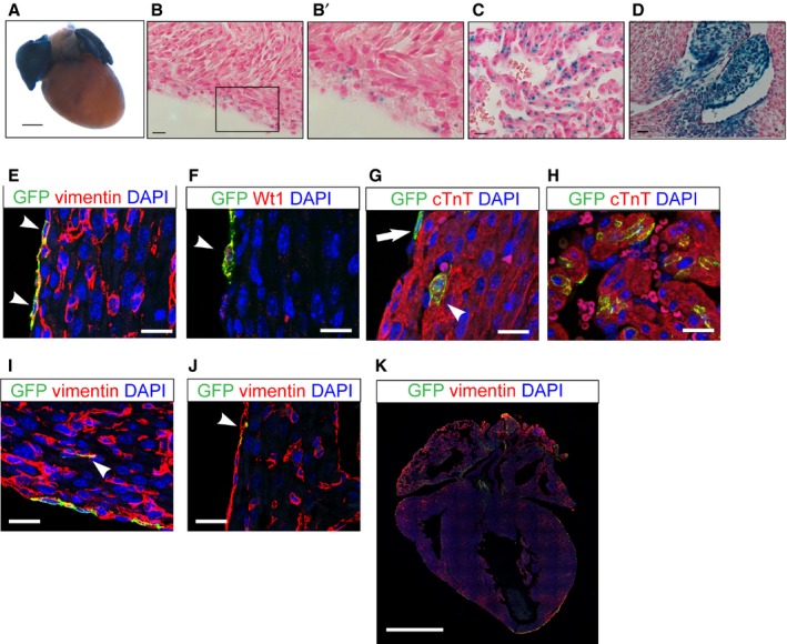
The neonatal heart atrial cardiomyocytes and epicardial cells are Wnt responsive. Neonatal heart of Axin2‐lacZ mice at P1. A, Whole‐mount X‐gal staining of the neonatal heart. Scale bar=0.5 mm. B, X‐gal staining of the epicardial and subepicardial space. B’, Inlet enlarged from (B). C, X‐gal staining of atrial cells. D, P1 neonatal heart of Axin2‐lacZ mice showing Wnt‐responsive cells of valves. B through D, Scale bar=10 μm. E through K, Immunofluorescent staining to characterize Axin2‐positive cells using Axin2‐CreERT2;mTmG neonatal heart labeled at P0 and traced for 2 days. E, Immunostaining against vimentin (red) and GFP (green) in the epicardium. Arrowheads indicate double‐positive cells. F, Immunostaining against Wilms tumor 1 (Wt1) and GFP. Arrowhead indicates a double‐positive cell. G, Immunostaining against cTnT and GFP in subepicardial cardiomyocytes. The arrow indicates a GFP‐positive epicardial cell not positive for cTnT. The arrowhead indicates a double‐positive cell. H, Immunostaining against cTnT and GFP in atria. I, Immunostaining against vimentin and GFP in the cardiac interstitium. Arrowhead indicates double‐positive interstitial cells. J, Immunostaining against vimentin and GFP in the endocardium. Arrowhead indicates double‐positive cells. E through J, Scale bar=20 μm. K, Immunostaining against vimentin and GFP in a lower magnification stitching image. Scale bar=1 mm. cTnT indicates cardiac troponin T; GFP, green fluorescent protein; P1, postnatal day 1.