
Figure 6.
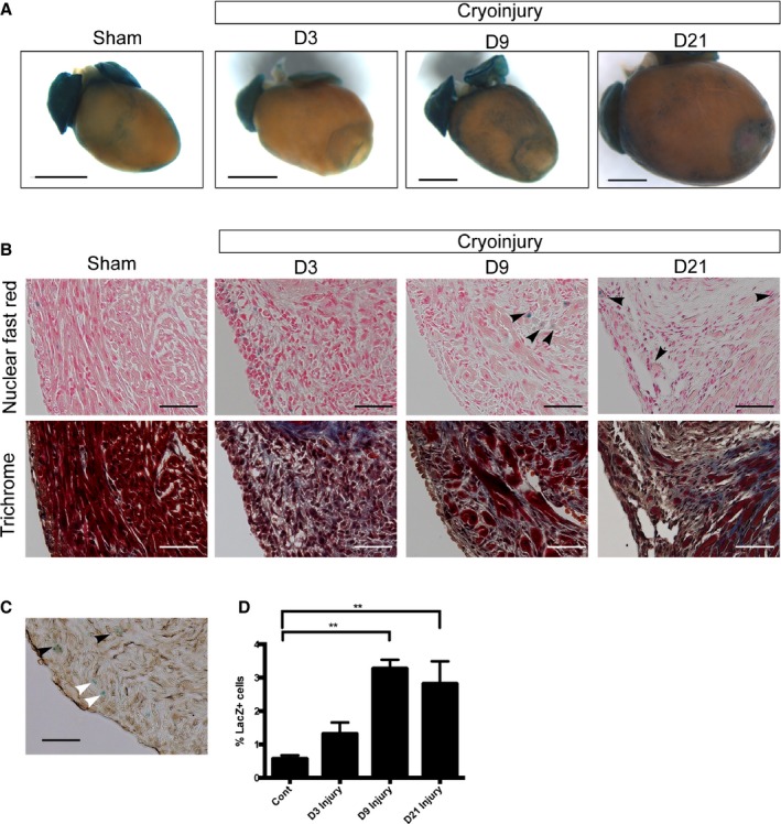
Activation of Wnt/β‐catenin signaling in the injured neonatal heart. A, Whole‐mount pictures of X‐gal–stained hearts of Axin2‐lacZ mice at 3, 9, and 21 days after injury. A whole‐mount picture of the X‐gal–stained sham‐operated heart at 3 days after surgery is also shown. Scale bar=1 mm. B, X‐gal and trichrome staining in serial sections showing the border area at 3, 9, and 21 days after injury and the equivalent area in the sham‐operated heart. Arrowheads indicate lacZ‐positive cells in the myocardium at 9 days after the injury. C, X‐gal and vimentin staining (brown) showing that some lacZ‐positive cells overlap (black arrowheads), whereas others do not (white arrowheads). D, Quantification of Axin2‐lacZ positive cells in the border area at 3, 9, and 21 days after injury and the equivalent area in the sham‐operated hearts at 3 days. Scale bar=100 μm. D indicates day. **denotes P<0.01.