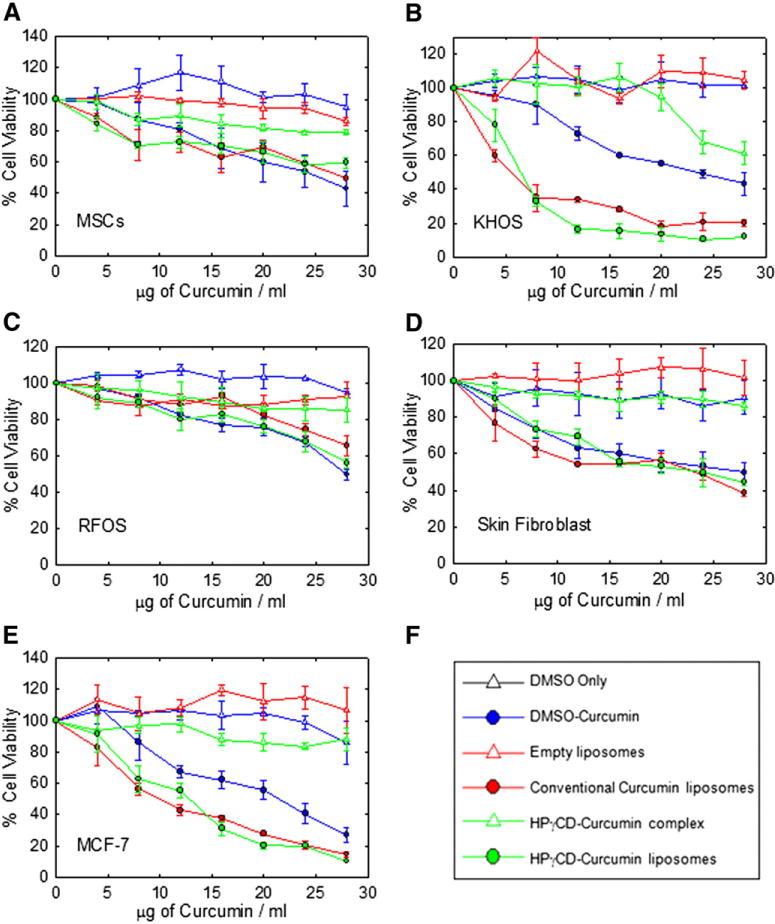
Figure 2.

Effect of different curcumin formulations on cell lines of mesenchymal and epithelial origin. (A) Human mesenchymal stem cells (B) KHOS (C) RFOS (D) Skin fibroblast (E) MCF-7 (F) Legends. Values represent the mean ± SD of triplicate experiments after 48 h of exposure to different curcumin formulations.
