
Fig. 2.
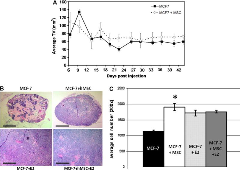
hMSCs promote tumor cell proliferation in the absence of estrogen. a Tumor volume in mm3 (mean ± SEM). About 4–6-week-old female ovariectomized SCID/beige mice injected subcutaneously with 1 × 10⁶ MCF-7 ± 1 × 10⁶ hMSC in 50 μl of sterile PBS with 100 μl reduced growth factor matrigel (BD biosystems), n = 5 mice per group. Tumors were measured every 3 days. b H&E sections at 509 showing differences in tumor cellularity. Scale bar equal to 500 μm. c Cell count averages per field of view at 200×. Bars represent average cell number ± SE, (*, P < 0.001)