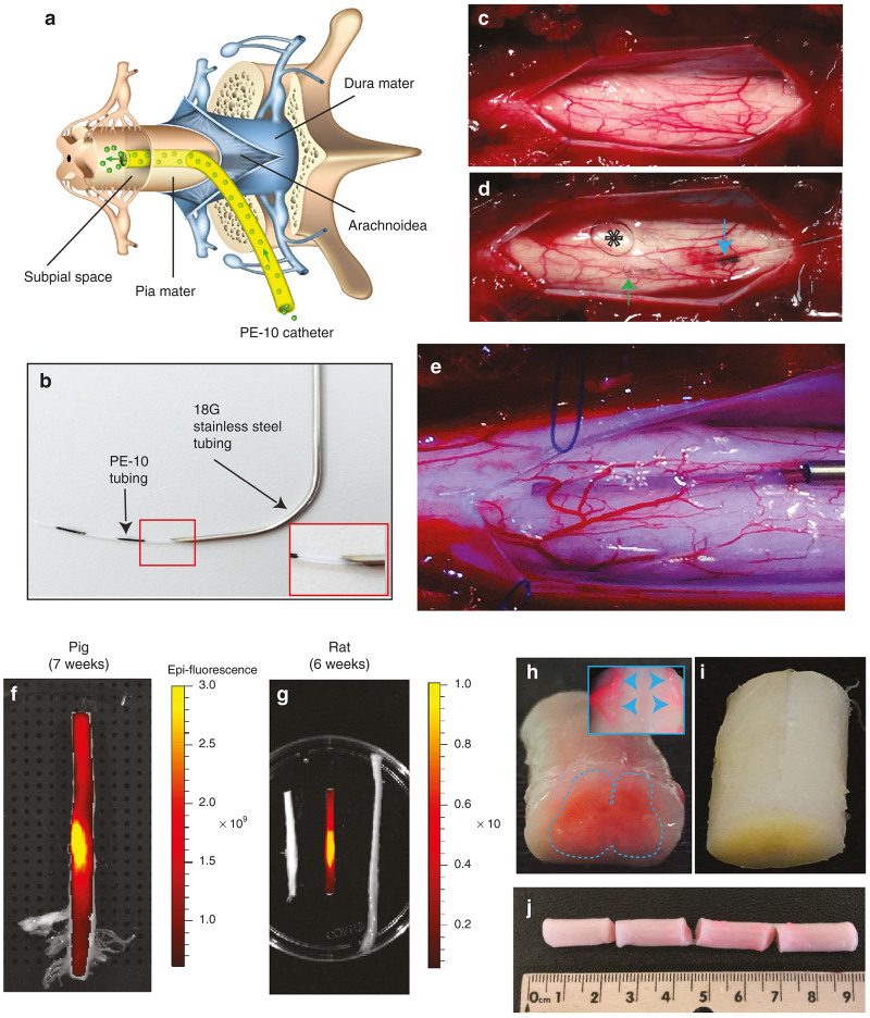
Figure 1.
Technique of subpial AAV9 delivery and macroscopically defined spinal cord surface transgene expression. (a) Schematic drawing of spinal cord, meninges and sub-pially placed PE-10 catheter in pigs. (b) A catheter guiding tube (18G) is used to advance the PE-10 catheter into the subpial space. (c–e) To place the catheter into the subpial space, the dura mater is first cut open (c) and the catheter is advanced into the SP space (d,e). An air bubble injected into the subpial space can be seen (d-asterisk). (f,g) Surface GFP fluorescence densitometry shows an intense signal in both rat and pig spinal cord with the most intense GFP fluorescence seen at the epicenter of lumbar subpial injection. (h–j) The presence of intense RFP fluorescence in the spinal cord parenchyma detected macroscopically in pig thoracic spinal cord (h,j). A clear high level of RFP expression in ventral roots can also be seen (h-insert). No fluorescence in the control noninjected spinal cord can be identified (i).

