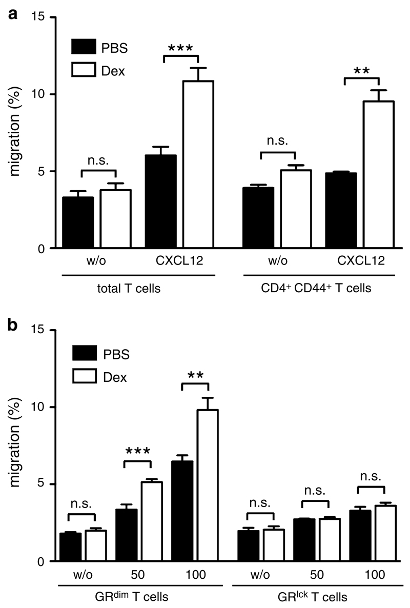
Fig. 4.
CXCL12-directed T cell migration is enhanced after Dex treatment. a Total T cells were isolated from spleens and lymph nodes of C57Bl/6 mice, pretreated for 3 h with or without Dex and tested in a transwell assay for their capacity to migrate towards 50 ng/ml mouse CXCL12 during a 3-h period without further presence of Dex. Cell numbers were determined by FACS using reference beads (left panel, N = 15). Results for CD44+ CD4+ T cells were calculated by FACS analyses of transwell assays using total T cells (right panel, N = 3). All values are depicted as mean ± SEM. Statistical analysis was performed using the unpaired t test. b Isolated T cells from spleens and lymph nodes of GRdim or GRlck mice, both on a C57Bl/6 background, were tested in a transwell assay for their capacity to migrate towards 50 or 100 ng/ml mouse CXCL12. The cells were pretreated for 3 h with or without Dex and subsequently allowed to migrate for another 3 h without further presence of Dex. Cell numbers were determined by FACS using reference beads (N = 6−8 for GRdim T cells, N = 10−12 for GRlck T cells). All values are depicted as mean ± SEM. Statistical analysis was performed using the unpaired t test

