Figure 2.
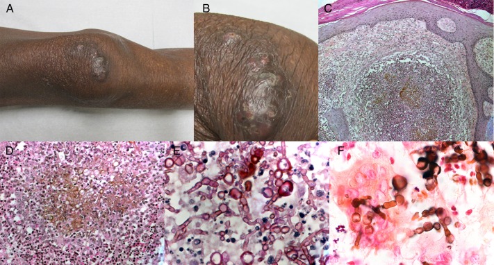
Patient 13. Pigmented infiltrated plaque of the right knee associated with diffuse subcutaneous infiltration in a 62-year-old kidney transplant recipient. (A) Full and (B) close-up views (courtesy of Camille Frances). (C and D) Hematoxylin-eosin staining revealed a dense dermal infiltrate with granulomatous inflammation, associating neutrophils, lymphocytes, epithelioid, and multinucleated cells, as well as pigmented fungal hyphae ([C], ×100; [D], ×400). (E) Periodic acid-Schiff staining showed septate fungal hyphae (×1000). (F) Fontana-Masson staining confirmed the pigmented character of fungal structures (×1000).

