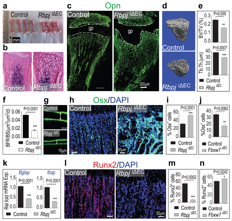
Figure 2. Endothelial Notch signalling regulates osteogenesis.
a, Decreased length of freshly dissected RbpjiΔEC femurs compared to control littermates.
b, Hematoxylin/eosin-stained longitudinal RbpjiΔEC and control tibia sections.
c, Osteopontin (Opn) immunostaining showing defective formation of trabeculae in P28 RbpjiΔEC tibia.
d, 3D µ-CT reconstruction of 4 week-old control and RbpjiΔEC metaphysis.
e, Reduced trabecular bone volume density measured as bone volume/total volume (BV/TV) and trabecular bone thickness (Tb.Th) in RbpjiΔEC mice.
f, g, Bone Formation Rate per Bone Surface (BFR/BS, f) calculated by calcein double labelling (7 day time interval) confirmed decreased bone formation in P28 Rbpj mutants. Arrows in (g) mark distance between calcein-labelled layers (n=10 mice from 6 independent litters). Error bars, ± s.e.m. P value, two-tailed unpaired t-tests.
h, Osterix (Osx) immunostaining shows strongly increased osteoprogenitor numbers in RbpjiΔEC metaphysis.
i, j, Quantitation of metaphyseal Osx+ cells in RbpjiΔEC (i) and Fbxw7iΔEC mutants (j) relative to controls (n=6 mice from 4 independent litters). Error bars, ± s.e.m. P values, two-tailed unpaired t-tests.
k, qPCR analysis showing reduced expression of mature osteoblast markers (Bglap, Ibsp) in RbpjiΔEC bones (n=6 mice from 4 independent litters).
1, Immunostaining showing decrease of Runx2+ early osteoprogenitors in 4 week-old RbpjiΔEC tibiae.
m, n, Quantitation of metaphyseal Runx2+ cells in RbpjiΔEC (m) and Fbxw7iΔEC mutants (n) relative to littermate controls (n=6 mice from 4 independent litters). Error bars, ± s.e.m. P values, two-tailed unpaired t-tests.

