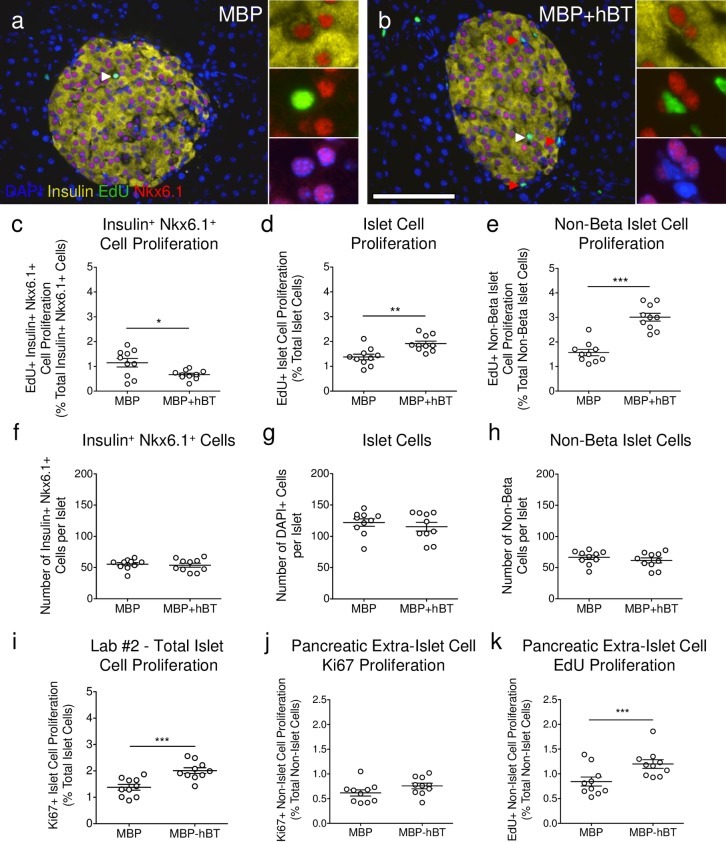
Fig 6. ANGPTL8 treatment in mice increases non-β-cell proliferation.
(a-b) Staining performed by Lab #2 for insulin (yellow), EdU (green), Nkx6.1 (red), and DAPI (blue) for (a) MBP and (b) MBP+hBT islets. White arrowheads indicate a proliferating insulin+ Nkx6.1+ DAPI+ cell [inset (a)] and red arrowheads indicate a proliferating insulin- Nkx6.1- DAPI+ cell [inset (b)]. Scale bar: 100 μm. (c-e) Quantification of EdU+ proliferation for (c) β-cells, (d) total islet cells, and (e) non-β islet cells. (f-h) Quantification of (f) β-cell number, (g) total islet cell number, and (h) non-β islet cell number per islet. Islet cells were identified by dilating insulin area by one cell’s diameter and filling all holes within the object. β-cells were identified by Nkx6.1+ cells co-localized with DAPI surrounded by insulin. Non-β islet cells were calculated by subtracting the β-cell counts from the total islet cell counts. (i) Total islet cell proliferation by Ki67 from original stained slides by Lab #2 examined in Fig 3. (j-k) Quantification of pancreatic proliferation by (j) Ki67+ or (k) EdU+ (% of total non-islet cells) from original stained slides by Lab #2 examined in Fig 3. Data are mean ± SEM. MBP and MBP+hBT = 10 animals per group. Student’s t test was performed. * p < 0.05, ** p < 0.01, *** p < 0.001.

