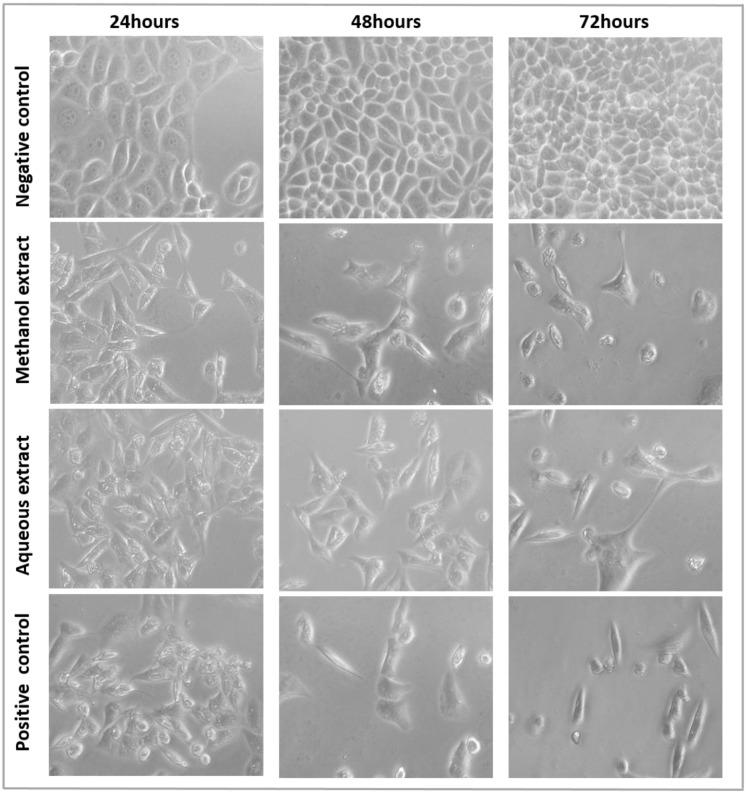
Fig 5. The morphological changes of MDA-MB-231 cells treated with methanolic and aqueous extracts at their respective IC50.
MDA-MB-231 cells treated with 8.5 μg/mL tamoxifen were used a positive control. After 24, 48, and 72 h of treatment, the cell morphological alterations were observed with an inverted-phase contrast microscope.

