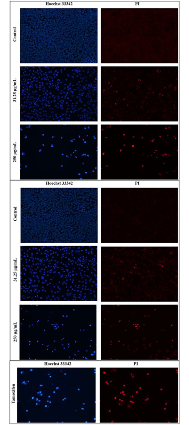
Fig 7. Fluorescence imaging for detection of apoptosis in MDA-MB-231 cells. Cells were treated with A) methanolic extract B) aqueous extract, and C) positive control (tamoxifen) for 24 h at 31.25 and 250 μg/mL.
Left panel displays Hoechst 33342 staining while right panel displays PI staining of the same field. The morphological alterations in cells were visualized under a fluorescence microscope (magnification: 200x). Extract-treated cells showed condensed and fragmented nuclei at both tested concentrations. The number of distorted nuclei and apoptotic cells were higher at 250 μg/mL than at 31.25 μg/mL. The percentage of apoptotic cells was higher among cells treated with the methanolic extract than among those treated with the aqueous extract.

